Article

Shock Management for Cardio-surgical Intensive Care Unit Patient: The Silver Days

Register or Login to View PDF Permissions
Permissions× For commercial reprint enquiries please contact Springer Healthcare: ReprintsWarehouse@springernature.com.

For permissions and non-commercial reprint enquiries, please visit Copyright.com to start a request.

For author reprints, please email rob.barclay@radcliffe-group.com.
Average (ratings)
No ratings
Your rating

Abstract

Shock in cardio-surgical intensive care unit (ICU) patients requires prompt identification of the underlying condition and timely therapeutic interventions. Management during the first 6 hours, also referred to as “the golden hours”, is of paramount importance to reverse the shock state and improve the patient’s outcome. The authors have previously described a state-of-the-art diagnostic work-up and discussed how to optimise preload, vascular tone, contractility, heart rate and oxygen delivery during this phase. Ideally, shock can be reversed during this initial period. However, some patients might have developed multiple organ dysfunction, which persists beyond the first 6 hours despite the early haemodynamic treatment goals having been accomplished. This period, also referred to as “the silver days”, is the focus of this review. The authors discuss how to reduce vasopressor load and how to minimise adrenergic stress by using alternative inotropes, extracorporeal life-support and short acting beta-blockers. The review incorporates data on fluid weaning, safe ventilation, daily interruption of sedation, delirium management and early rehabilitation. It includes practical recommendations in areas where the evidence is scarce or controversial. Although the focus is on cardio-surgery ICU patients, most of the considerations apply to critical ill patients in general.

Disclosure:The authors have no conflicts of interest to declare.

Received:

Accepted:

Correspondence Details:A Rudiger, Institute of Anesthesiology, University Hospital Zurich, Raemistrasse 100, CH-8091 Zurich, Switzerland. E: alain.rudiger@usz.ch

Copyright Statement:

The copyright in this work belongs to Radcliffe Medical Media. Only articles clearly marked with the CC BY-NC logo are published with the Creative Commons by Attribution Licence. The CC BY-NC option was not available for Radcliffe journals before 1 January 2019. Articles marked ‘Open Access’ but not marked ‘CC BY-NC’ are made freely accessible at the time of publication but are subject to standard copyright law regarding reproduction and distribution. Permission is required for reuse of this content.

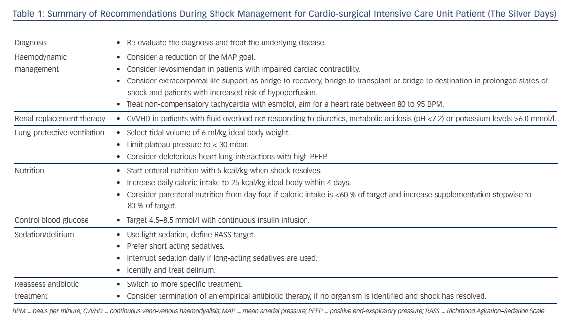
Shock in cardio-surgical intensive care unit (ICU) patients is a serious condition associated with a high morbidity and mortality.1,2 Prompt identification of the underlying condition and timely therapeutic interventions are key to reverse the shock state and to improve the patients’ outcome. Hence, the management during the first 6 hours is of paramount importance. This time period is also referred to as “the golden hours”. Ideally, a correct diagnosis is established allowing specific treatments. The authors have previously described a state-ofthe-art diagnostic work-up and discussed how to optimise preload, vascular tone, contractility, heart rate and oxygen delivery during this phase.3 Ideally, shock can be reversed during this initial period, however some patients might have developed multiple organ dysfunction that persists beyond the first 6 hours despite the early haemodynamic treatment goals been accomplished.4 This period, also referred to as “the silver days”, is the focus of this review. The authors discuss the management of organ dysfunction in critically ill patients after cardiac surgery. The following recommendations (summarised in Table 1) are not exclusive, rather they highlight some important considerations to be made while treating these patients after the initial resuscitation phase.

Methods

For this narrative review, a search of the PubMed database and a review of bibliographies from selected articles were performed to identify original data relating to this topic. Key words used for the search were, among others, “haemodynamic management” “vasopressors”, “levosimendan”, inotropes”, inotropic therapy”, “nutrition”, “sedation”, “ventilation”, “weaning from mechanical ventilation”, “delirium” and “procalcitonin”. National and international guidelines were reviewed and integrated, e.g. the “Guidelines for the Diagnosis and Treatment of Acute and Chronic Heart Failure” of the European Society of Cardiology (ESC)5 and the “Consensus on circulatory shock and haemodynamic monitoring“ from the Task force of the Society of Intensive Care Medicine (ESICM).6

Articles were scrutinised regarding their study design, population evaluated, interventions, outcomes and limitations. Finally, personal recommendations were included and highlighted as such to give a comprehensive overview on this topic.

Optimise Haemodynamics

Reduce Vasopressor Load

The optimal mean arterial blood pressure (MAP) under vasopressor therapy is still under investigation. For patients with septic shock, no benefit was found after increasing the MAP by use of vasopressors above 65–70 mmHg.7 Moreover, higher vasopressor loads were associated with higher mortality.8 However Asfar et al. showed that targeting a MAP of 80–85 mmHg in patients with chronic arterial hypertension reduced the incidence of renal replacement therapy.7 Therefore, the authors follow the recommendations of the 2014 consensus report of the ESICM6 and target an individualised blood pressure rather than fixed MAP-goals after the first phase of lifesaving measures. In patients that remain anuric despite a MAP of 70 mmHg, the blood pressure target is reduced. In patients with critical vascular stenosis or right heart failure, the MAP is not reduced <65 mmHg. In all others, MAP targets are continuously reduced and sometimes tolerated as low as 50 mmHg in order to reduce the amount of vasopressors necessary.

Table 1: Summary of Recommendations During Shock Management for Cardio-surgical Intensive Care Unit Patient (The Silver Days)

Article image

Consider Alternative Inotropes

Beta-adrenergic drugs have been associated with considerable risks including adverse effects on metabolism, bacterial growth and alterations of the innate immune response.9–13 Also, the amount and duration of catecholamines is independently associated with adverse cardiac events such as tachyarrhythmia and prolonged elevated heart rate.14 Hence, efforts are necessary to limit the use of these drugs as much as possible. One alternative is the inodilator levosimendan. It increases the troponin C affinity for Ca2+, which results in strengthening of the myocardial contraction without increasing oxygen demand.15 Also, it has vasodilatorproperties via activation of ATP-dependent potassium channels.16 The use of Levosimendan in cardiogenic shock is controversial because patients with cardiogenic shock have been excluded in safety studies as levosimendan causes vasodilation.17 It is recommended by the 2013 ESC guidelines on heart failure in cardiogenic shock and second line treatment for low output heart failure if the effect of beta blockage is thought to be the reason for hypoperfusion.5 The Levosimendan Infusion versus Dobutamine (LIDO) study showed a survival benefit of levosimendan after 6 months.16,18 The Survival Of Patients With Acute Heart Failure In Need Of Intravenous Inotropic Support (SURVIVE) study found an advantage in patients with decompensated chronic heart failure previously treated with beta blockers.17,19,20 Another study showed the combination of levosimendan and dobutamine to be more effective compared to dobutamine alone.21 In cardiac surgery, levosimendan has shown to produce a dose dependent effect on stroke volume, also when given preoperatively in patient at risk, and to shorten length of ICU stay.22 It is the only inotrope which might decrease the mortality after cardiac surgery.23

The authors use levosimendan for persisting heart failure after shock resolution, weaning failure from inotropic therapy, or weaning from extracorporeal life support (ECLS) with a dose of 0.1–0.2 mcg/kg/min without a bolus, if systolic blood pressure is >100 mmHg with low dose vasopressors (noradrenaline <0.1 mcg/kg/min). Experimental and clinical studies also suggest an improvement of cardiac function with levosimendan in patients with septic shock.17,24

Evaluate Mechanical Support

In situations with increasing inotrope requirements or inadequate oxygen delivery despite high doses of inotropes, mechanic circulatory support must be evaluated.25 ECLS with an extracorporeal membrane oxygenator can be established quickly in experienced hands and offers an opportunity for temporary haemodynamic support.26 This might give time for decision-making (bridge to decision) or for the ventricle to recover (bridge to recovery). The ESC guidelines give IIb and IIa recommendations for short-term mechanical support as a bridge to decision and bridge to recovery, respectively.5 In patients with multiple organ dysfunctions mortality rates may be excessively high, prohibiting the use of ECLS in this particular patient population.27 Recently, the concept of awake ECLS has been introduced.28 The benefits of having patients on ECLS without mechanical ventilation include the possibility to assess the patients’ cognitive functions. It also allows interaction with the patient and the possibility to inquire his will, particularly in view of future therapeutic options (ventricular assist device, transplantation). As positive pressure ventilation and sedation can be avoided, patients awake on ECLS have a better haemodynamic stability.

In selected patients with persisting cardiogenic shock under optimised pharmacologic therapy and/or weaning failure from ECLS, commercially available left- or biventricular assist devices (LVAD or BVAD) will provide sufficient organ perfusion for everyday life. These devices may be used in end-stage heart failure patients before heart transplantation (bridge to transplantation).26 Mechanical support will allow early rehabilitation leading to improved nutritional state, muscle strength and physical performance status. According to the ESC heart-failure guidelines, LVADs are a IIa indication for long-term use when transplantation is not possible (bridge to destination).5

Advantages and Risks of Extracorporeal Life Support

Article image

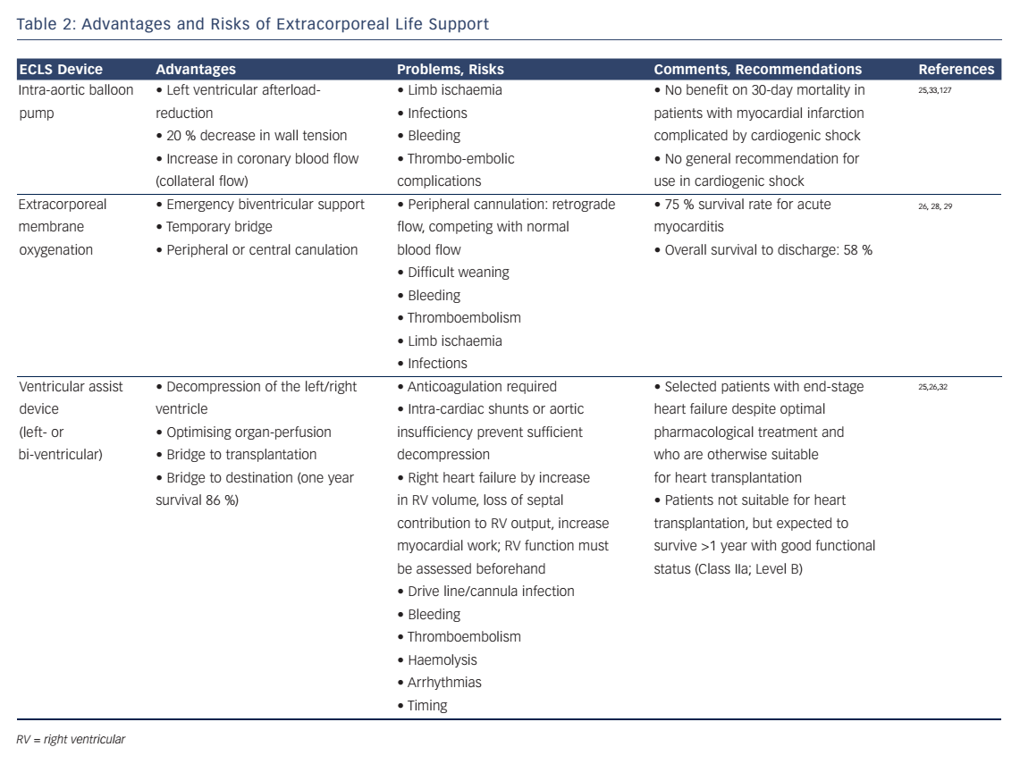
Increased cardiac output by a LVAD may enlarge right ventricular (RV) volume load and cause RV-failure. Therefore, RV function must be assessed before considering the implantation of a LVAD.25 Surely the decision should be made in a multidisciplinary approach by experts due to the invasive nature of these devices. Complications may be related to the mandatory use of anticoagulation (bleeding, thromboembolism), mechanical shear stress on cellular blood components (haemolysis) and long-term infections of the implanted materials (drive line, cannulas, device).25,26,29–33 A summary of the advantages and risks of ECLS is provided in Table 2.

Control Heart Rate

Adrenergic stress might induce inflammation and contribute to the pathogenesis of organ dysfunction.12,34 Hence, the use of beta-adrenergic drugs must be limited to a minimum. Taking this concept to the next level, the theoretical benefits of beta-blockers during critical illness have been discussed.35 Recently, Morelli et al. tested the effects of esmolol in septic patients.36 He included high-risk patients, who required noradrenaline and had a heart rate ≥95 beats per minute (BPM) despite 24 hours of haemodynamic optimisation (MAP ≥65 mmHg, pulmonary artery occlusion pressure ≥ 12 mmHg, SvO2 ≥65 %). Esmolol was started at a dose of 0.5 mg/min and increased by 0.5–1.0 mg/min increments at 20-minute intervals to an upper dose limit of 30 mg/min. The goal was a heart rate reduction to a rate between 80 to 94 BPM. If mixed venous oxygen saturation (SmvO2) decreased below 65 % and/or arterial lactate concentrations increased despite appropriate oxygenation (SaO2 ≥95 %) and a haemoglobin concentration ≥8 g/dl, levosimendan was administered at a dose of 0.05–0.2 mcg/kg/min for 24 hours. In their cohort study from 2013, the authors showed improvements in stroke volume, a decrease in norepinephrine requirement and a reduction of mortality.36 If these results are confirmed in future multicentre trials, this concept might revolutionise current practice in sepsis management. Potentially, this new concept might be usefully expanded to other critical ill patients with non-compensatory tachycardia.

A reduction in heart rate will allow a better ventricular filling in patients with diastolic myocardial dysfunction, resulting in an increase in stroke volume. The same is true for patients with supraventricular arrhythmias and atrial fibrillation (AF) with a high ventricular response rate.37 Additionally, a reduction in heart rate will reduce myocardial oxygen expenditure, hence inducing cardio-protection. However, some patients will have a high heart rate in order to compensate for a low stroke volume and/or an insufficient oxygen delivery. In these patients, prolonged heart rate reduction will result in haemodynamic collapse and eventually death. Hence, short-acting drugs should be used in patients in which it is unclear whether tachycardia is compensatory or not. In such situations, the authors use esmolol, a selective beta1- blocker with a short half-life of 9 minutes, as an intravenous bolus (10–20 mg-wise up to 1 mg/kg), followed by a continuous infusion of 0.05 mg/kg/min. The infusion rate can be increased every 30 minutes if needed. Of note, the negative inotropic effects of esmolol must be balanced against the potential benefits, and close haemodynamic monitoring including echocardiography is mandatory in unstable patients treated with beta-blockers.

Start Fluid Weaning

Fluid resuscitation is one of the corner stones of shock therapy to restore tissue perfusion.38 However, it was demonstrated that a liberal fluid regime increased mortality and morbidity in a diverse group of patients.39 Targeting a central venous pressure (CVP) between 8–12 mmHg seemed to impair the microcirculation, was a risk factor for acute kidney injury (AKI) and increased mortality.39 The underlying mechanism seems to be a consecutive reduction in renal blood flow and glomerular filtration rate by a high venous pressure.39,40 Of note, there is no good correlation between CVP and fluid responsiveness41–43 and therefore other preload variables should be used.44

Since fluid overload leads to increased morbidity in acute respiratory distress syndrome,45 pancreatitis46 and sepsis,47 the authors recommend a conservative fluid regime with fluids being administered preferably after assessing responsiveness.6 As soon as the patient has stabilised and noradrenaline requirements are below 10 mcg/min, fluid weaning is started. In haemodynamic stabile patients with AKI, protocolled renal replacement therapy (RRT) allows large negative fluid balances.48 Net fluid removal usually causes improvements in lung function, therefore leading to a reduction in ventilation days. Reduction of oedema in the bowel and the extremities will facilitate nutrition and mobilisation of the patient.

Ventilate Safely

The authors use also the concept of lung-protective ventilation derived from acute respiratory distress syndrome (ARDS) patients for ICU patients with normal lungs. A tidal volume of ≤6 ml/kg should be chosen with plateau pressure of <30 mbar. Positive end-expiratory pressures (PEEP) are increased with increasing oxygen requirements. However, high PEEP levels increase RV afterload and might cause RV dysfunction.49 These heart-lung interactions might reduce RV stroke volume and consequently cardiac output, thereby decreasing oxygen delivery to the periphery. In case of further aggravation of hypoxaemia under increased PEEP, a persisting foramen ovale should be excluded. Hypercarbia causes pulmonary vasoconstriction and increases RV afterload, thus normoventilation should be targeted if possible in patients with pulmonary artery hypertension and/or RV dysfunction. Another risk of invasive ventilation is ventilator associated pneumonia (VAP), which was defined by the following criteria during a time frame of 48 hours after intubation:50,51

  • new or progressive pulmonary infiltrates;
  • fever;
  • leucocytosis;
  • purulent secretion;
  • reduction of the PaO2:FiO2 ratio by ≥ 15 %.

In the American Thoracic Society definition from 2005, VAP is defined as pneumonia occuring 48–72 hours after intubation.52 However, diagnosis of VAP remains difficult as criteria are nonspecific and symptoms overlap as in conditions like sepsis, ARDS or atelectasis.50 Radiographic signs are neither specific nor sensitive. In a study comparing chest X-ray findings in autopsy proven pneumonia, no sign had a diagnostic efficiency greater than 68 % and positive air bronchogramms predicted 65 % of pneumonias.53 Thus, a diagnosis of VAP is made by a combination of clinical signs, radiographic findings and, importantly, microbiological cultures.54,55

To reduce the incidence of VAP our hospital implemented the following bundle:56

  • Strict hand disinfection,
  • oral hygiene with application of chlorhexidin,
  • elevation of upper body >30° (if possible from a haemodynamic point of view),
  • defined weaning and sedation protocols,
  • continous subglottical suction,
  • cuffpressure-control,
  • daily reevaluation of gastric ulcer prophylaxis,
  • Routine change of ventilator circuits and filters.

Manage Acute Renal Dysfunction

Understand Renal Dysfunction

Acute kidney injury (AKI) has a high incidence in patients after cardiac surgery varying between 1 and 30 %.57–61 The acute kidney injury network (AKIN) and risk, injury, failure, loss (RIFLE) criteria of AKI are accurate and early predictive of mortality with creatinine levels being the most useful marker.57,59,62,63 The pathophysiology is complex and involves pre-, intra- and post-operative risk factors.57,64 Most vulnerable is the patient with pre-existing renal dysfunction prior to surgery.57,59 Nephrotoxins such as contrast agents and antibiotics, impaired cardiac output and poor renal perfusion, inflammation caused by surgical trauma, cardio-pulmonary-bypass and sepsis are further factors contributing to AKI.59,65 No preventive pharmacological therapy has been proven to be effective although studies indicate a possible benefit of statins pre-operatively.57,66–68 If possible, cardiac surgery should be performed ≥24 hours after coronary angiography to reduce the risk of AKI.69 Patients who need renal replacement therapy have a 27 fold increased mortalitiy.70

Start Renal Replacement Therapy

The ideal time point of RRT-initiation remains unclear since the benefits of RRT must outweigh the risks of RRT61,71–74 In their cardiosurgical ICU the authors start RRT with continuous veno-venous haemodialysis (CVVHD), if the patient has metabolic acidosis (pH <7.2), increasing potassium levels (>6.0 mmol/l), complications of azotaemia (blood urea >20 mmol/l) such as encephalopathy or fluid overload not responding to diuretics.

The current Kidney Disease Improving Global Outcomes (KDIGO) guidelines recommend a dose of 20–25 ml/kg/hour of effluent flow in patients with AKI emphasising the need to individualise the patients dose by assessing volume status, acid-base status and electrolyte disturbances.62 Anticoagulation is required to prevent membrane clotting and dysfunction. Regional anticoagulation with citrate has the advantage over heparin75 to reduce the bleeding risk in post-operative patients after cardiac surgery, as heparin anticoagulation can be complicated by platelet- and red blood cell-consumption.75 However, impaired cellular aerobic metabolism (Krebs cycle) puts the patients at risk for insufficient citrate metabolism and citrate accumulation. In order to avoid this risk, the authors accept post-filter levels of calcium [ionised] as high as 0.5 mmol/l and limit the amount of citrate used (blood flow as low as 100 ml/minute, citrate concentration fix 3 mmol/l). The calcium quotient (total calcium [albumin corrected] divided by calcium [ionised] is measured daily. A value of ≥2.5 points to citrate accumulation and should prompt either a reduction of citrate load or a switch to alternative anticoagulation strategies.

Interrupt Sedation Daily

After cardiac surgery most patients arrive in the ICU sedated and with some neuromuscular blockade. After resolving of neuromuscular blockade sedation should keep the patient pain-free but interactive, calm but lucid and cooperative.76 Over-sedation was associated with prolonged mechanical ventilation and ICU-stay, while light sedation was associated with reduced length of ICU and hospital stay, less post-traumatic stress disorders and improved survival rates.76–78

In order to minimise sedation, the authors use short-acting sedative agents such as propofol or dexmedetomidine (if tolerated from a haemodynamic point of view). A non-benzodiazepine approach in critically ill patients led to a shorter duration of mechanical ventilation, a reduced length of ICU stay79 and a lower mortality80 compared to the use of benzodiazepines. The benefits of dexmedetomidine include the better sleep architecture,81 a lower risk of delirium and a shorter length of stay after cardiac surgery.82 When long-active sedatives such as midazolam must be used due to haemodynamic instability, daily interruption of sedation is mandatory, as this has been shown to reduce days on the ventilator, ICU length of stay and even mortality.78,83

Detect and Treat Delirium

There is extensive evidence that delirium prolongs ICU stay84 and increases morbidity and mortality85–88 after cardiac surgery. Also a longer duration of delirium is associated with worse global cognition after discharge.89 Risk factors for delirium after cardiac surgery include age, preexisting cognitive impairment and cerebrovascular disease, benzodiazepine use and immobilsation.90 A review also pointed out that blood transfusion, mechanical ventilation and even use of intraaortic balloon pump91 are associated with increased risk of delirium. The confusion assessment method for the ICU (CAM-ICU) 92,93 and the intensive care delirium screening checklist (ICDSC) 94 are tools for the diagnosis of delirium in critically ill patients with reported pooled sensitivities and specificities of 80 % and 75 % for the ICDSC and 76 % and 96 % for the CAM-ICU.95 Scarce data exist for the treatment of delirium.88 Cooperative patients with a hypoactive form of delirium and/or hallucinations are treated with haloperidol, preferably orally and with low doses. In the authors’ institution, agitated and noncooperative patients are treated primarily with enteral pipamperon, a mild neuroleptic agent with sedative properties. The drug is usually given in the afternoon and evening (e.g. 4pm, 6pm and 8pm) to treat agitation and to induce sleep. Intravenous dexmedetomidine is added in severely agitated patients.

Rehabilitate Early

Prolonged critical illness results in loss of lean body mass and muscle weakness.96,97 Catecholamines induce myocyte apoptosis, and muscle weakness is potentially related to excessive sympathetic tone.12,98 Physiotherapy in the ICU is safe99 and can be started in cardiac surgery patients considering safety issues which are related to devices, sheaths or sternotomy.100 Early mobilisation and physiotherapy improves functional outcome and probably reduces length of ICU stay.101–103 Even in patients with ECLS, early physiotherapy and mobilisation is possible and safe.104,105

Feeding and Support Metabolism

Enteral Versus Parenteral Nutrition

Early enteral nutrition is recommended to preserve gastro-intestinal integrity and prevent bacterial translocation.106 On the other hand, enteral nutrition bears the risk of vomiting and aspiration, gastrointestinal obstruction and bowel ischaemia. This is particularly true during prolonged shock, when blood is redistributed from the gut to vital organs such as the brain and the heart.

The authors start nutrition with 5 kcal/kg/day after shock resolution (normalised lactate levels, decreasing noradrenaline requirements) and increase stepwise to 25 kcal/kg/day over the next days. In patients without a contraindication for enteral nutrition, an early initiation of parenteral nutritision caused longer ICU stays and higher incidence of ICU aquired infections and higher health care costs.107 Other complications such as poor glycaemic control were described with parenteral nutrition.108,109 Uncertainty exists in patients with contrandications for enteral feeding: a meta-analysis of older studies (between 1981 and 1994) showed an association with higher infection rates in patients with parenteral nutrition,110 whereas a newer study showed a shorter duration of mechanical ventilation (without effect on 60-day mortality) if patients received parenteral nutrition within 24 hours of ICU admission.111

Recently, a study showed benefits of parenteral nutrition was started on day four if enteral intake was <60 % of the targeted calories.112 Therefore, the authors start parenteral nutrition between days four and eight if shock has resolved and enteral calory intake is <60 % of the targeted calories. As with enteral nutrition, parenteral nutrition is gradually established over days, up to 80 % of the target calory intake.

Stress Ulcer Prophylaxis

Following international guidelines, the authors recommend stress ulcer prophylaxis with proton pump inhibitors (e.g. entral or intravenous pantoprazole 40 mg once daily) in patients with risk factor of gastro-intestinal bleeding (coagulopathy or anticoagulation, prolonged mechanical ventilation >48 hours, hypotension, steroid therapy).113 Meta analysis showed significantly less upper gastrointestinal bleeding with prophylaxis (in the absence of any mortality benefit).114–116 It is worth noting that stress ulcer prophylaxis is a grade 2C recommendation in the 2013 surviving sepsis campaign.113 Since there is an increased risk of pneumonia with increased stomach pH in ambulant patients,117 there might be a greater incidence of VAP with use of stress ulcer prophylaxis. Clostridium difficile infections have also be associated with the use of prophylaxis.118 Taking these considerations into account, the use of stress ulcer prophylaxis should be limited to patients at risk of bleeding, until enteral nutrition is fully established.

Blood Glucose Control

Hyperglycaemia is common in patients with shock due to the physiological stress reaction. Excessive glucose plasma levels have been associated with adverse outcome.119,120 However, pharmacological glucose control bears the risk of hypoglycaemia.121 A large, international randomised trial showed that a liberal glucose management (targeting glucose levels <10 mmol/l) resulted in a lower mortality than targeting a glucose level of 4.5–6 mmol/l.122 As numerous arterial blood gas analyses including glucose measurements are performed, the authors have a low rate of accidental hypoglycemic episodes. In their ICU, the authors have agreed on blood glucose targets between 4.5–8.5 mmol/l using continuous insulin infusions.

Use DVT Prophylaxis

As critical ill patients are at risk for deep vein thrombosis (DVT),113,123 prophylaxis is warranted. The decision when to start and how to provide DVT prophylaxis may be difficult as the bleeding risk is high after cardiac surgery. In a prospective multicentre trial independent predictors for major bleeding in patients receiving heparin thrombosis prophylaxis were described, including renal replacement therapy, low platelet count and antiplatelet agents during the past 7 days.124

The authors usually start DVT prophylaxis with a continuous infusion of 10,000 IE/24h of unfractionated heparin 6 hours after surgery. In patients with an indication for therapeutic anticoagulation, the heparin dose is increased by 2,000 to 5,000 IE every 6 hours until the anti-Xa activity is between 0.3 and 0.7 IE/ml.

Infection Control

During the early phase of shock, broad-spectrum intravenous antibiotics are used if sepsis is suspected and after samples for microbiology have been taken. Importantly, antibiotic therapy has to be adapted according to the microbiology results, or even stopped if the patient improves and no pathogen is identified.113

The use of procalcitonin (PCT) as a marker to guide duration of antibiotic therapy reduces length and cost of antibiotic therapy.125 However, PCT levels usually increase after cardiac surgery even without infections.126 PCT should therefore not be used as a sole marker of infection to guide antibiotic therapy in patients after cardiac surgery.

Conclusion

Shock in cardio-surgical patients is a life-threatening disorder. Multiple organ failure can occur despite early diagnosis and prompt therapeutic interventions. The period beyond the first 6 hours of shock onset, also referred to as the silver days, is the focus of the current review. The authors discuss haemodynamic management, ventilation strategies, renal replacement therapy, sedation and nutrition in affected patients. Over the years, allegedly life-saving treatments were shown to have rather harmful effects in critically ill patients, particularly if prolonged use or high dosages are applied. Potentially harmful interventions include haemodynamic optimisation to supranormal targets, excessive fluid administration, over-feeding and oversedation. Hence, treatments of patients during the silver days must focus on reducing harm and avoiding complications related to critical care.4,127 This concept includes avoidance of fluid overload, reduction of excessive catecholamines, safe ventilation and daily interruption of sedation. Although the authors focus on cardio-surgery ICU patients, most of the considerations apply to critical-ill patients in general.

References

  1. Mazzeffi M, Zivot J, Buchman T, et al. In-hospital mortality after cardiac surgery: patient characteristics, timing, and association with postoperative length of intensive care unit and hospital stay. Ann Thorac Surg 2014;97:1220–5.
    Crossref | PubMed
  2. Hsu P-S, Chen J-L, Hong G-J, et al. Extracorporeal membrane oxygenation for refractory cardiogenic shock after cardiac surgery: predictors of early mortality and outcome from 51 adult patients. Eur J Cardiothorac Surg 2010;37:328–33.
    Crossref | PubMed
  3. Hauffe T, Krüger B, Bettex D, et al. Shock management for cardio-surgical ICU patients – the golden hours. Card Fail Rev 2015;1 :75–82.
    Crossref 
  4. Marshall JC. Critical illness is an iatrogenic disorder. Crit Care Med 2010;38:S582–9.
    Crossref | PubMed
  5. McMurray JJ, Adamopoulos S, Anker SD, et al. ESC Guidelines for the diagnosis and treatment of acute and chronic heart failure 2012: The Task Force for the Diagnosis and Treatment of Acute and Chronic Heart Failure 2012 of the European Society of Cardiology. Developed in collaboration with the Heart Failure Association (HFA) of the ESC. Eur Heart J 2012;33:1787–847. 
    Crossref | PubMed
  6. Cecconi M, De Backer D, Antonelli M, et al. Consensus on circulatory shock and hemodynamic monitoring. Task force of the European Society of Intensive Care Medicine. Intensive Care Med 2014:1795–815. PMCID: PMC4239778
    Crossref | PubMed
  7. Asfar P, Meziani F, Hamel J-F, et al. High versus low bloodpressure target in patients with septic shock. N Engl J Med 2014;370:1583–93. 
    Crossref | PubMed
  8. Dünser MW, Ruokonen E, Pettilä V, et al. Association of arterial blood pressure and vasopressor load with septic shock mortality: a post hoc analysis of a multicenter trial. Crit Care 2009;13:R181. PMCID: PMC2811945
    Crossref | PubMed
  9. Lyte M, Freestone PPE, Neal CP, et al. Stimulation of Staphylococcus epidermidis growth and biofilm formation by catecholamine inotropes. Lancet (London, England) 2003;361 :130–135.
    Crossref | PubMed
  10. Freestone PPE, Haigh RD, Lyte M. Specificity of catecholamine-induced growth in Escherichia coli O157:H7, Salmonella enterica and Yersinia enterocolitica. FEMS Microbiol Lett 2007;269:221–228.  
    Crossref | PubMed
  11. Singer M. Catecholamine treatment for shock-equally good or bad? Lancet 2007;370:636–637.  
    Crossref | PubMed
  12. Dünser MW, Hasibeder WR. Sympathetic overstimulation during critical illness: adverse effects of adrenergic stress. J Intensive Care Med 2009;24:293–316. 
    Crossref | PubMed
  13. Hartemink KJ, Johan Groeneveld a. B. Vasopressors and inotropes in the treatment of human septic shock: Effect on innate immunity? Inflammation 2012;35:206–213. PMCID: PMC3282003
    Crossref | PubMed
  14. Schmittinger C a, Torgersen C, Luckner G, et al. Adverse cardiac events during catecholamine vasopressor therapy: a prospective observational study. Intensive Care Med 2012;38:950–8.  
    Crossref | PubMed
  15. Papp Z, Édes I, Fruhwald S, et al. Levosimendan: molecular mechanisms and clinical implications: consensus of experts on the mechanisms of action of levosimendan. Int J Cardiol 2012;159:82–7.
    Crossref | PubMed
  16. Pathak A, Lebrin M, Vaccaro A, et al. Pharmacology of levosimendan: inotropic, vasodilatory and cardioprotective effects. J Clin Pharm Ther 2013;38:341–9. 
    Crossref | PubMed
  17. Pierrakos C, Velissaris D, Franchi F, et al. Levosimendan in critical illness: a literature review. J Clin Med Res 2014;6:75–85. PMCID: PMC3935527
    Crossref | PubMed
  18. Follath F, Cleland J, Just H, et al. Efficacy and safety of intravenous levosimendan compared with dobutamine in severe low-output heart failure (the LIDO study): a randomised double-blind trial. Lancet 2002;360:196–202.
    Crossref | PubMed
  19. Mebazaa A, Nieminen MS, Packer M, et al. Levosimendan vs dobutamine for patients with acute decompensated heart failure: the SURVIVE randomized trial. JAMA 2007;297:1883–91.  
    Crossref | PubMed
  20. Mebazaa A, Nieminen MS, Filippatos GS, et al. Levosimendan vs. dobutamine: outcomes for acute heart failure patients on beta-blockers in SURVIVE. Eur J Heart Fail 2009;11 :304–11. PMCID: PMC2645051
    Crossref | PubMed
  21. Nanas JN, Papazoglou PP, Terrovitis J V, et al. Hemodynamic effects of levosimendan added to dobutamine in patients with decompensated advanced heart failure refractory to dobutamine alone. Am J Cardiol 2004;94:1329–32.
    Crossref | PubMed
  22. Toller W, Heringlake M, Guarracino F, et al. Preoperative and perioperative use of levosimendan in cardiac surgery: European expert opinion. Int J Cardiol 2015;184:323–36. 
    Crossref | PubMed
  23. Landoni G, Biondi-Zoccai G, Greco M, et al. Effects of levosimendan on mortality and hospitalization. A metaanalysis of randomized controlled studies. Crit Care Med 2012;40:634–46. 
    Crossref | PubMed
  24. Rudiger A, Singer M. The heart in sepsis: from basic mechanisms to clinical management. Curr Vasc Pharmacol 2013;11 :187–95.
    Crossref | PubMed
  25. Lighthall GK, Singh S. Perioperative maintenance of tissue perfusion and cardiac output in cardiac surgery patients. Semin Cardiothorac Vasc Anesth 2014;18:117–36. 
    Crossref | PubMed
  26. Peura JL, Colvin-Adams M, Francis GS, et al. Recommendations for the use of mechanical circulatory support: device strategies and patient selection: a scientific statement from the American Heart Association. Circulation 2012;126:2648–67. 
    Crossref | PubMed
  27. Schmidt M, Burrell A, Roberts L, et al. Predicting survival after ECMO for refractory cardiogenic shock: the survival after veno-arterial-ECMO (SAVE)-score. Eur Heart J 2015;36:2246–56.
    Crossref | PubMed
  28. Sommer W, Marsch G, Kaufeld T, et al. Cardiac awake extracorporeal life support-bridge to decision? Artif Organs 2015;39:400–8. 
    Crossref | PubMed
  29. Luo X, Wang W, Hu S, et al. Extracorporeal membrane oxygenation for treatment of cardiac failure in adult patients. Interact Cardiovasc Thorac Surg 2009;9:296–300. 
    Crossref | PubMed
  30. Pages ON, Aubert S, Combes A, et al. Paracorporeal pulsatile biventricular assist device versus extracorporal membrane oxygenation-extracorporal life support in adult fulminant myocarditis. J Thorac Cardiovasc Surg 2009;137:194–7. 
    Crossref | PubMed
  31. Thiele H, Ohman EM, Desch S, et al. Management of cardiogenic shock. Eur Heart J 2015;36:1223–3.
    Crossref | PubMed
  32. Vincent JL, De Backer D. Circulatory shock. N Engl J Med 2013;369:1726–34.  
    Crossref | PubMed
  33. Birati EY, Rame JE. Left ventricular assist device management and complications. Crit Care Clin 2014;30:607–27. 
    Crossref | PubMed
  34. Singer M. Catecholamine treatment for shock – equally good or bad? Lancet 2007;370:636–7. 
    Crossref | PubMed
  35. Rudiger A. Beta-block the septic heart. Crit Care Med 2010;38:S608–12. 
    Crossref | PubMed
  36. Morelli A, Ertmer C, Westphal M, et al. Effect of heart rate control with esmolol on hemodynamic and clinical outcomes in patients with septic shock: a randomized clinical trial. JAMA 2013;310:1683–91.
    Crossref | PubMed
  37. Arrigo M, Bettex D, Rudiger A. Management of atrial fibrillation in critically ill patients. Crit Care Res Pract 2014:840615. PMCID: PMC3914350
    Crossref | PubMed
  38. Myburgh JA, Mythen MG. Resuscitation fluids. N Engl J Med 2013;369:1243–51. 
    Crossref | PubMed
  39. Marik PE. Iatrogenic salt water drowning and the hazards of a high central venous pressure. Ann Intensive Care 2014;4:21. PMCID: PMC4122823
    Crossref | PubMed
  40. Prowle JR, Kirwan CJ, Bellomo R. Fluid management for the prevention and attenuation of acute kidney injury. Nat Rev Nephrol 2014;10:37–47.
    Crossref | PubMed
  41. Marik PE, Cavallazzi R. Does the central venous pressure predict fluid responsiveness? An updated meta-analysis and a plea for some common sense. Crit Care Med 2013;41 :1774–81. 
    Crossref | PubMed
  42. Marik PE, Baram M, Vahid B. Does central venous pressure predict fluid responsiveness? A systematic review of the literature and the tale of seven mares. Chest 2008;134:172–8. 
    Crossref | PubMed
  43. Cecconi M, Aya HD. Central venous pressure cannot predict fluid-responsiveness. Evid Based Med 2014;19:63.
    Crossref | PubMed
  44. Cecconi M, Hofer C, Teboul J-L, et al. Fluid challenges in intensive care: the FENICE study: a global inception cohort study. Intensive Care Med 2015;41 :1529–37. PMCID: PMC4550653
    Crossref | PubMed
  45. Gattinoni L, Cressoni M, Brazzi L. Fluids in ARDS: from onset through recovery. Curr Opin Crit Care 2014;20:373–7. 
    Crossref | PubMed
  46. de-Madaria E, Soler-Sala G, Sánchez-Payá J, et al. Influence of fluid therapy on the prognosis of acute pancreatitis: a prospective cohort study. Am J Gastroenterol 2011;106:1843–50. 
    Crossref | PubMed
  47. Boyd JH, Forbes J, Nakada T, et al. Fluid resuscitation in septic shock: a positive fluid balance and elevated central venous pressure are associated with increased mortality. Crit Care Med 2011;39:259–65. 
    Crossref | PubMed
  48. Ganter CC, Hochuli R, Bossard M, et al. Forced fluid removal in critically ill patients with acute kidney injury. Acta Anaesthesiol Scand 2012;56:1183–90. 
    Crossref | PubMed
  49. Jardin F, Vieillard-Baron A. Is there a safe plateau pressure in ARDS? The right heart only knows. Intensive Care Med 2007;33:444–7. 
    Crossref | PubMed
  50. Choudhuri AH. Ventilator-associated pneumonia: when to hold the breath? Int J Crit Illn Inj Sci 2013;3:169–74. PMCID: PMC3883194
    Crossref | PubMed
  51. Safdar N, Dezfulian C, Collard HR, et al. Clinical and economic consequences of ventilator-associated pneumonia: a systematic review. Crit Care Med 2005;33:2184–93. 
    Crossref | PubMed
  52. Guidelines for the management of adults with hospitalacquired, ventilator-associated, and healthcare-associated pneumonia. Am J Respir Crit Care Med 2005;171 :388–416. 
    Crossref | PubMed
  53. Wunderink RG, Woldenberg LS, Zeiss J, et al. The radiologic diagnosis of autopsy-proven ventilator-associated pneumonia. Chest 1992;101 :458–63. 
    Crossref | PubMed
  54. Klompas M, Kleinman K, Platt R. Development of an algorithm for surveillance of ventilator-associated pneumonia with electronic data and comparison of algorithm results with clinician diagnoses. Infect Control Hosp Epidemiol 2008;29:31–7.
    Crossref | PubMed
  55. Lalwani S, Mathur P, Tak V, et al. Diagnosis of ventilatorassociated pneumonia: comparison between ante-mortem and post-mortem cultures in trauma patients. Indian J Med Microbiol 2014;32:294–300.  
    Crossref | PubMed
  56. Sommerstein R, Nemeth J, Braun A Von. Pneumonie beim Erwachsenen. Schweiz Med Forum 2014;14:590–3.
  57. Kramer RS, Herron CR, Groom RC, et al. Acute kidney injury subsequent to cardiac surgery. J Extra Corpor Technol 2015;47:16–28.  PMCID: PMC4566816
    PubMed
  58. Szelkowski LA, Puri NK, Singh R, Massimiano PS. Current trends in preoperative, intraoperative, and postoperative care of the adult cardiac surgery patient. Curr Probl Surg 2015;52:531–69.  
    Crossref | PubMed
  59. Rosner MH. Acute kidney injury associated with cardiac surgery. Clin J Am Soc Nephrol 2005;1 :19–32.
    Crossref | PubMed
  60. Huen SC, Parikh CR. Predicting acute kidney injury following cardiac surgery: a systematic review. Ann Thorac Surg 2013;93:337–47. PMCID: PMC3286599
    Crossref | PubMed
  61. Villa G, Ricci Z, Ronco C. Renal replacement therapy. Crit Care Clin 2015;31 :839–48.
    Crossref | PubMed
  62. Kellum JA, Lameire N. Diagnosis, evaluation, and management of acute kidney injury: a KDIGO summary (Part 1). Crit Care 2013;17:204; PMCID: PMC4057151
    Crossref | PubMed
  63. Shann KG, Likosky DS, Murkin JM, et al. An evidence-based review of the practice of cardiopulmonary bypass in adults: a focus on neurologic injury, glycemic control, hemodilution, and the inflammatory response. J Thorac Cardiovasc Surg 2006;132:283–90. 
    Crossref | PubMed
  64. Bellomo R, Kellum JA, Ronco C. Acute kidney injury. Lancet (London, England) 2012;380:756–66.
    Crossref | PubMed
  65. Rudiger A, Singer M. Acute kidney injury. Lancet 2012;380:1904; author reply 1905. 
    Crossref | PubMed
  66. Tabata M, Khalpey Z, Pirundini PA, et al. Renoprotective effect of preoperative statins in coronary artery bypass grafting. Am J Cardiol 2007;100:442–4. 
    Crossref | PubMed
  67. Virani SS, Nambi V, Polsani VR, et al. Preoperative statin therapy decreases risk of postoperative renal insufficiency. Cardiovasc Ther 2010;28:80–6. 
    Crossref | PubMed
  68. Song Y, Kwak YL, Choi YS, et al. Effect of preoperative statin therapy on myocardial protection and morbidity endpoints following off-pump coronary bypass surgery in patients with elevated C-reactive protein level. Korean J Anesthesiol 2010;58:136–41. PMCID: PMC2872864
    Crossref | PubMed
  69. Ranucci M, Ballotta A, Agnelli B, et al. Acute kidney injury in patients undergoing cardiac surgery and coronary angiography on the same day. Ann Thorac Surg 2013;95:513–9.
    Crossref | PubMed
  70. Chertow GM, Levy EM, Hammermeister KE, et al. Independent association between acute renal failure and mortality following cardiac surgery. Am J Med 1998;104:343–8 
    Crossref | PubMed
  71. Gaffney AM, Sladen RN. Acute kidney injury in cardiac surgery. Curr Opin Anaesthesiol 2015;28:50–59.
    Crossref | PubMed
  72. Liu Y, Davari-Farid S, Arora P, et al. Early versus late initiation of renal replacement therapy in critically ill patients with acute kidney injury after cardiac surgery: a systematic review and meta-analysis. J Cardiothorac Vasc Anesth 2014;28:557–63.
    Crossref | PubMed
  73. Karvellas CJ, Farhat MR, Sajjad I, et al. A comparison of early versus late initiation of renal replacement therapy in critically ill patients with acute kidney injury: a systematic review and meta-analysis. Crit Care 2011;15:R72. PMCID: PMC3222005
    Crossref | PubMed
  74. Wang X, Jie Yuan W. Timing of initiation of renal replacement therapy in acute kidney injury: a systematic review and meta-analysis. Ren Fail 2012;34:396–402. 
    Crossref | PubMed
  75. Bonassin Tempesta F, Rudiger A, Previsdomini M, et al. Platelet consumption and filter clotting using two different membrane sizes during continuous venovenous haemodiafiltration in the intensive care unit. Crit Care Res Pract 2014;2014:203637.  PMCID: PMC4020532
    Crossref | PubMed
  76. Reade MC, Finfer S. Sedation and delirium in the intensive care unit. N Engl J Med 2014;370:444–54. 
    Crossref | PubMed
  77. Gradwohl-Matis I, Mehta S, Dünser MW. What’s new in sedation strategies? Intensive Care Med 2015;102:1696–9.
    Crossref | PubMed
  78. Kress JP, Pohlman AS, O’Connor MF, et al. Daily interruption of sedative infusions in critically ill patients undergoing mechanical ventilation. N Engl J Med 2000;342:1471–7. 
    Crossref | PubMed
  79. Fraser GL, Devlin JW, Worby CP, et al. Benzodiazepine versus nonbenzodiazepine-based sedation for mechanically ventilated, critically ill adults: a systematic review and metaanalysis of randomized trials. Crit Care Med 2013;41 :S30–8. 
    Crossref | PubMed
  80. Lonardo NW, Mone MC, Nirula R, et al. Propofol is associated with favorable outcomes compared with benzodiazepines in ventilated intensive care unit patients. Am J Respir Crit Care Med 2014;189:1383–94. 
    Crossref | PubMed
  81. Jakob SM. Dexmedetomidine vs midazolam or propofol for sedation during prolonged mechanical ventilation. JAMA 2012;307:1151.
    Crossref | PubMed
  82. Lin YY, He B, Chen J, et al. Can dexmedetomidine be a safe and efficacious sedative agent in post-cardiac surgery patients? a meta-analysis. Crit Care 2012;16:R169. PMCID: PMC3682268
    Crossref | PubMed
  83. Girard TD, Kress JP, Fuchs BD, et al. Efficacy and safety of a paired sedation and ventilator weaning protocol for mechanically ventilated patients in intensive care (awakening and breathing controlled trial): a randomised controlled trial. Lancet (London, England) 2008;371 :126–34. 
    Crossref | PubMed
  84. Norkiene I, Ringaitiene D, Misiuriene I, et al. Incidence and precipitating factors of delirium after coronary artery bypass grafting. Scand Cardiovasc J 2007;41 :180–5. 
    Crossref | PubMed
  85. Clarke SP, McRae ME, Del Signore S, et al. Delirium in older cardiac surgery patients. J Gerontol Nurs 2010;58:7250–7. 
    Crossref | PubMed
  86. Saczynski JS, Marcantonio ER, Quach L, et al. Cognitive trajectories after postoperative delirium. N Engl J Med 2012;367:30–39. PMCID: PMC3433229
    Crossref | PubMed
  87. Rudolph JL, Inouye SK, Jones RN, et al. Delirium: an independent predictor of functional decline after cardiac surgery. J Am Geriatr Soc 2010;58:643–9. PMCID: PMC2856754
    Crossref | PubMed
  88. Jackson P, Khan A. Delirium in critically Ill Patients. Crit Care Clin 2015;31 :589–603.
    Crossref | PubMed
  89. Pandharipande PP, Girard TD, Jackson JC, et al. Long-term cognitive impairment after critical illness. N Engl J Med 2013;369:1306–16. PMCID: PMC3922401
    Crossref | PubMed
  90. McPherson JA, Wagner CE, Boehm LM, et al. Delirium in the cardiovascular ICU: exploring modifiable risk factors. Crit Care Med 2013;41 :405–13. PMCID: PMC3557701
    Crossref | PubMed
  91. Katznelson R, Djaiani GN, Borger MA, et al. Preoperative use of statins is associated with reduced early delirium rates after cardiac surgery. Anesthesiology 2009;110:67–73.
    Crossref | PubMed
  92. Ely EW, Inouye SK, Bernard GR, et al. Delirium in mechanically ventilated patients: validity and reliability of the confusion assessment method for the intensive care unit (CAM-ICU). JAMA 2001;286:2703–10. 
    Crossref | PubMed
  93. Ely EW, Margolin R, Francis J, et al. Evaluation of delirium in critically ill patients: validation of the confusion assessment method for the intensive care unit (CAM-ICU). Crit Care Med 2001;29:1370–9. 
    Crossref | PubMed
  94. Bergeron N, Dubois MJ, Dumont M, et al. Intensive care delirium screening checklist: evaluation of a new screening tool. Intensive Care Med 2001;27:859–64.  
    Crossref | PubMed
  95. Neto AS, Nassar AP, Cardoso SO, et al. Delirium screening in critically ill patients: a systematic review and metaanalysis. Crit Care Med 2012;40:1946–51.  
    Crossref | PubMed
  96. Vanhorebeek I, Van den Berghe G. The neuroendocrine response to critical illness is a dynamic process. Crit Care Clin 2006;22:1–15. 
    Crossref | PubMed
  97. Batt J, dos Santos CC, Cameron JI, et al. Intensive care unit– acquired weakness. Am J Respir Crit Care Med 2013;187:238–46. 
    Crossref | PubMed
  98. Ng Y, Goldspink DF, Burniston JG, et al. Characterisation of isoprenaline myotoxicity on slow-twitch skeletal versus cardiac muscle. Int J Cardiol 2002;86:299–309. 
    Crossref | PubMed
  99. Parker A, Sricharoenchai T, Needham DM. Early rehabilitation in the intensive care unit: preventing physical and mental health impairments. Curr Phys Med Rehabil Rep 2013;1 :307–14. PMCID: PMC3889146
    Crossref | PubMed
  100. Hodgson CL, Stiller K, Needham DM, et al. Expert consensus and recommendations on safety criteria for active mobilization of mechanically ventilated critically ill adults. Crit Care 2014;18:658. PMCID: PMC4301888
    Crossref | PubMed
  101. Stiller K. Physiotherapy in intensive care: an updated systematic review. Chest 2013;144:825–47.
    Crossref | PubMed
  102. Schweickert WD, Pohlman MC, Pohlman AS, et al. Early physical and occupational therapy in mechanically ventilated, critically ill patients: a randomised controlled trial. Lancet 2009;373:1874–82. 
    Crossref | PubMed
  103. Morris PE, Goad A, Thompson C, et al. Early intensive care unit mobility therapy in the treatment of acute respiratory failure. Crit Care Med 2008;36:2238–43.
    Crossref | PubMed
  104. Freeman R, Maley K. Mobilization of intensive care cardiac surgery patients on mechanical circulatory support. Crit Care Nurs Q 2013;36:73–88. 
    Crossref | PubMed
  105. Ko Y, Cho YH, Park YH, et al. Feasibility and safety of early physical therapy and active mobilization for patients on extracorporeal membrane oxygenation. ASAIO J 2015:564–8.
    Crossref | PubMed
  106. Kreymann KG, Berger MM, Deutz NEP, et al. ESPEN Guidelines on enteral nutrition: Intensive care. Clin Nutr 2006;25:210–23. 
    Crossref | PubMed
  107. Casaer MP, Mesotten D, Hermans G, et al. Early versus late parenteral nutrition in critically ill adults. N Engl J Med 2011;365:506–17.
    Crossref | PubMed
  108. Mongardon N, Singer M. The evolutionary role of nutrition and metabolic support in critical illness. Crit Care Clin 2010;26:443–50.
    Crossref | PubMed
  109. Malone AM. Permissive underfeeding: its appropriateness in patients with obesity, patients on parenteral nutrition, and non-obese patients receiving enteral nutrition. Curr Gastroenterol Rep 2007;9:317–22. 
    Crossref | PubMed
  110. Braunschweig CL, Levy P, Sheean PM, et al. Enteral compared with parenteral nutrition: a meta-analysis. Am J Clin Nutr 2001;74:534–42. 
    PubMed
  111. Doig GS, Simpson F, Sweetman EA, et al. Early parenteral nutrition in critically ill patients with short-term relative contraindications to early enteral nutrition: a randomized controlled trial. JAMA 2013;309:2130–8
    Crossref | PubMed
  112. Heidegger CP, Berger MM, Graf S, et al. Optimisation of energy provision with supplemental parenteral nutrition in critically ill patients: a randomised controlled clinical trial. Lancet 2013;381 :385–93.
    Crossref | PubMed
  113. Dellinger RP, Levy MM, Rhodes A, et al. Surviving sepsis campaign: international guidelines for management of severe sepsis and septic shock: 2012. Crit Care Med 2013;41 :580–637. 
    Crossref | PubMed
  114. Kahn JM, Doctor JN, Rubenfeld GD. Stress ulcer prophylaxis in mechanically ventilated patients: integrating evidence and judgment using a decision analysis. Intensive Care Med 2006;32:1151–8. 
    Crossref | PubMed
  115. Cook DJ, Reeve BK, Guyatt GH, et al. Stress ulcer prophylaxis in critically ill patients. Resolving discordant meta-analyses. JAMA 275:308–14. 
    Crossref | PubMed
  116. Marik PE, Vasu T, Hirani A, et al. Stress ulcer prophylaxis in the new millennium: a systematic review and metaanalysis. Crit Care Med 2010;38:2222–8. 
    Crossref | PubMed
  117. Laheij RJF, Sturkenboom MCJM, Hassing R-J, et al. Risk of community-acquired pneumonia and use of gastric acidsuppressive drugs. JAMA 2004;292:1955–60.
    Crossref | PubMed
  118. Plummer MP, Blaser AR, Deane AM. Stress ulceration: prevalence, pathology and association with adverse outcomes. Crit Care 2014;18:213. PMCID: PMC4056012
    Crossref | PubMed
  119. Furnary AP, Wu Y. Clinical effects of hyperglycemia in the cardiac surgery population: the portland diabetic project. Endocr Pract 12 Suppl 3:22–6. 
    Crossref | PubMed
  120. Desai SP, Henry LL, Holmes SD, et al. Strict versus liberal target range for perioperative glucose in patients undergoing coronary artery bypass grafting: a prospective randomized controlled trial. J Thorac Cardiovasc Surg 2012;143:318–25.
    Crossref | PubMed
  121. Gandhi GY. Intensive intraoperative insulin therapy versus conventional glucose management during cardiac surgery. Ann Intern Med 2007;146:233. 
    Crossref | PubMed
  122. Finfer S, Chittock DR, Su SY-S, et al. Intensive versus conventional glucose control in critically ill patients. N Engl J Med 2009;360:1283–97.
    Crossref | PubMed
  123. Cade JF. High risk of the critically ill for venous thromboembolism. Crit Care Med 1982;10:448–50.
    Crossref | PubMed
  124. Lauzier F, Arnold DM, Rabbat C, et al. Risk factors and impact of major bleeding in critically ill patients receiving heparin thromboprophylaxis. Intensive Care Med 2013;39:2135–43. 
    Crossref | PubMed
  125. Heyland DK, Johnson AP, Reynolds SC, et al. Procalcitonin for reduced antibiotic exposure in the critical care setting: a systematic review and an economic evaluation. Crit Care Med 2011;39:1792–9.
    Crossref | PubMed
  126. Sponholz C, Sakr Y, Reinhart K, et al. Diagnostic value and prognostic implications of serum procalcitonin after cardiac surgery: a systematic review of the literature. Crit Care 2006;10:R145.  PMCID: PMC1751067
    Crossref | PubMed
  127. Singer M, Glynne P. Treating Critical Illness: The importance of first doing no harm. PLoS Med 2005;2:e167. PMCID: PMC1160576
    Crossref | PubMed